STED microscopy
STED microscopy (STimulated Emission Depletion) is a super-resolution imaging technique based for resolving objects smaller than the diffraction-limit. Like conventional confocal microscope, the STED microscope acquires images by point scanning over the sample. But unlike confocal microscope, a second donut shaped depletion laser beam is aligned temporally and spatially with the normal excitation laser such that a specific emission of fluorescent dyes occurs within a central [very small 20-80 nm diameter] region in the sample only. In doing so, this STED laser essentially reduces the point spread function (PSF) to produce a crisp image.
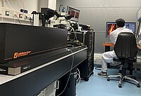
Our new STED microscope is a custom-built one from Abberior. It is an upright microscope equipped with several state-of-the-art technologies and modalities.
The system is equipped with advanced adoptive optics (AO) such as deformable mirror so as to reduce aberrations, and to improve image quality by manipulating the optical wavefront.
Similarly, the objective is mounted on a z-piezo fast objective scanner which enables us to take all kinds of 2-D images (X/Y, X/Z, Y/Z) as well as 3-D images (x/z/y-stacks). With this technology, biological structures can be physically imaged in a defined 2-D plane which otherwise is only possible via orthogonal projection during 3D- image post-processing.
The image acquisition is conducted via Imspector software which is again equipped with most advanced imaging packages/tools such as Easy3D, RESCue, DyMIN, and MINFIELD to generate the high-resolution image while reducing photobleaching at the same time.
Features
Excitation system
Epifluorescence illumination 405/470/590/635nm
Four fluorescence excitation lasers (405nm, 488nm, 518nm, 561nm, 640nm)
Two STED lasers (595nm, 775nm)
Titanium-sapphire laser for multi-photon imaging
Microscope and objectives
BX61Wl-F microscope upright body
Fixed stage
100X, NA 1.4 oil Immersion objective lens
60X NA 1.3 Silicon Immersion objective lens
60X NA 1.2 water dipping long working distance objective lens
Detectors
2 spectral detection units with 2 APD detectors
PMT detector for reflection measurements
Software package
Imspector for data acquisition and data analysis
Easy3D for both 595 nm STED and 775 nm STED
RESCue, DyMIN, and MINFIELD imaging modes (package of three) for light dose reduction with STED and essential in live cell/tissue imaging.
Contact
Dr. Surya Rai, Tel.: +49-451-31017212, E-Mail: surya.rai(at)uni-luebeck.de
Selected publication
Wenzel, J., Lampe, J., Müller-Fielitz, H., Schuster, R., Zille, M., Müller, K., Krohn, M., Körbelin, J., Zhang, L., Özörhan, Ü., Neve, V., Wagner, J.U.G., Bojkova, D., Shumliakivska, M., Jiang, Y., Fähnrich, A., Ott, F., Sencio, V., Robil, C., Pfefferle, S., Sauve, F., Fernando Ferreira Coelho, C., Franz, J., Spieker, F., Lembrich, B., Binder, S., Feller, N., König, P., Busch, H., Collin, L., Villasenor, R., Jöhren, O., Altmeppen, H.C., Pasparakis, M., Dimmeler, S., Cinatl, J., Püschel, K., Zelic, M., Ofengeim, D., Stadelmann, C., Trottein, F., Nogueiras, R. Hilgenfeld, R., Glatzel, M., Prevot, V., Schwaninger, M. The SARS-CoV-2 main protease Mpro causes microvascular brain pathology by cleaving NEMO in brain endothelial cells. Nat Neurosci (2021) 24: 1522-1533

- Forschung
- Bridging Brain Barriers
- Cellular Electrophysiology
- Cerebral Perfusion and Metabolism
- Influence of RAAS on the Metabolic Syndrome
- Interaction between Tanycytes and the hormone axes
- Neuropeptides and Energy Homeostasis
- Neuroplasticity and Rhythms
- Pharmacovigilance
- WATCH group
- STED microscopy
- Publications
- Memberships
- Seminars
- Awards
- Technologies/Resources